
Molecular pathology of thymoma and thymic carcinoma

Abstract
Thymic epithelial tumors (TETs) comprise a heterogeneous group of epithelial-derived thymic neoplasms with diverse clinical behavior and underlying molecular genetic features. Owing to their rare nature, the molecular classification of TETs has only recently begun to be fully explored. The advent of advanced molecular studies, particularly the ability to sequence the DNA and RNA of tumors in a massively parallel fashion, has led to an increased understanding of the molecular underpinnings of thymic neoplasia. Thymomas, characterized by a heterogeneous group of molecular alterations, tend to have low mutational burdens and various copy number abnormalities including a characteristic loss of chromosomal material in the region of 6q25.2-p25.3, a recurrent, specific point mutation GTF2I p.L424H, and specific expression of certain microRNAs. Thymic carcinomas, in contrast, are generally characterized by increased tumor mutational burdens, multiple copy number alterations, and varied, non-recurrent, somatic mutations. Advances in molecular knowledge of TETs allow for more precise molecular classification of these tumors, and the presence of specific alterations aids in the diagnosis of borderline lesions. In the future, additional molecular studies will better delineate the molecular landscape of these tumors and may one day allow for more targeted treatment algorithms. This review aims to cover the current understanding of the molecular alterations thus far identified in thymomas and thymic carcinomas.
Keywords
INTRODUCTION
Thymic epithelial tumors (TETs) comprise a heterogeneous group of epithelial-derived thymic neoplasms with diverse clinical behavior and underlying molecular genetic features[1]. Thymomas and thymic carcinomas are the most common group of epithelial tumors arising within the mediastinum and represent a rare occurrence relative to other human cancers. They are estimated to have an incidence of 1.3 per 1,000,000 person-years, with only approximately 400 new cases diagnosed per year in the United States[2]. Thymomas make up the majority of lesions, with thymic carcinomas occurring only rarely. Even less commonly, epithelial-derived tumors with neuroendocrine differentiation or salivary gland type features may occur within the mediastinal compartment[3].
Traditionally and in current clinical practice, TETs are classified primarily based on histologic features. Numerous classification systems have been proposed and used over the years, with the World Health Organization (WHO) classification system being the most widely accepted[1,4-8]. Despite the presence of numerous histology-based classification systems, problems still exist with the most current classification schema due to the varied biological behavior of TETs[4-9]. It is known that in some instances, the clinical and biological behavior may differ between tumors with similar histology[9]. In addition, problems with interobserver reproducibility lead to difficulties in classifying tumors and predicting their biological behavior.
Staging of TETs is somewhat more simplified; the most commonly used staging is the modified Masaoka staging system, although more recently, a tumor-nodes-metastases system has been adopted by the American Joint Committee on Cancer in the 8th edition of their cancer staging manual[10-12]. However, even within the confines of these consolidated staging systems, tumors within the same stage category may still sometimes display varied biological behavior[13].
Owing to their rare nature, the molecular classification of TETs has only recently begun to be fully explored. The advent of advanced molecular studies, particularly the ability to sequence the DNA and RNA of tumors in a massively parallel fashion, has led to an increased understanding of the molecular underpinnings of thymic neoplasia. The varied biologic behavior of thymic epithelial neoplasms, including histologic classification and staging discrepancies, is likely explained by their underlying molecular features. This review evaluates the currently known molecular information, primarily focusing on thymomas and thymic carcinomas, and describes some of the evolving molecular classifications of these tumors that may one day lead to an integrated morpho-molecular classification that will better prognosticate these tumors and allow for potential future targeted therapies.
MOLECULAR FEATURES OF THYMOMA AND THYMIC CARCINOMA
Molecular landscape of thymoma
Thymomas are rare tumors that occur predominantly in the 5th and 6th decades of life, with an estimated incidence of approximately 15-30 cases per 100,000 people per year[14,15]. Currently, thymomas are classified based primarily on their histomorphologic features. The most commonly used classification system is that of the WHO, which classifies thymomas into five distinct subtypes based on the type of cell (spindle vs. epithelioid) and the relative proportion of associated lymphocytes[1]. The five distinct subtypes include type A (predominantly spindled), type AB (spindled with increased lymphocytes), type B1 (sparse epithelioid cells with abundant lymphocytes), type B2 (increased epithelioid cells with relatively fewer lymphocytes), and type B3 thymoma (epithelioid cells with atypical features and few lymphocytes) [Figure 1][2]. It is worth noting that this letter-number classification was originally meant to simply serve as a method for translating the prior Bernatz and Muller-Hermelink classification and lends itself to problems with reproducibility and is generally insufficient at classifying many of the distinct subtypes of thymoma that exist. Type A and AB tumors tend to present in lower clinical stages as compared to B1/B2/B3 thymomas[16]. A unique clinical characteristic of these tumors is their association with paraneoplastic syndromes, the most common of which is myasthenia gravis. Myasthenia gravis may occur in up to 25%-45% of thymoma patients and more commonly occurs in patients with lymphoid-rich tumors such as AB and B types[17]. Various other paraneoplastic syndromes including various immunodeficiency and autoimmunity states have been described and may be related to the underlying molecular biology of these tumors[18-20].
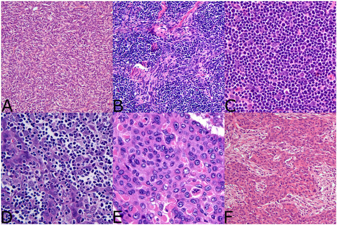
Figure 1. WHO histologic classification of thymoma (A) Type A thymoma is characterized by a spindle cell proliferation with limited numbers of lymphocytes, (B) Type AB thymoma is characterized by a spindle cell population intimately admixed with increased numbers of lymphocytes, (C) Type B1 thymoma is characterized by sparse epithelioid cells in a dense immature lymphoid cell population, (D) Type B2 thymoma is characterized by an increased number of epithelioid cells with a relatively decreased proportion of lymphoid cells, (E) Atypical thymoma (WHO type B3 thymoma) is characterized by sheets of epidermoid appearing epithelioid cells with increased cytologic atypia and few lymphocytes, (F) Metaplastic thymoma is characterized by a pseudosarcomatous stromal component with an admixed epithelial component composed of neoplastic thymic epithelial cells. Note: Not shown here are numerous other subtypes of more esoteric thymomas that do not fit into any particular classification system and have limited documented molecular data.
Owing to their rarity, the molecular characteristics of thymoma have lagged behind other more common tumor types. Currently, prognostication is based primarily upon post-surgical staging of the tumors; however, the lack of a more refined classification and prognostication system leads to difficulty in the management and treatment of patients with thymoma. In the past few decades, numerous studies have begun unraveling the molecular landscape of thymomas. Inoue et al.[21] performed an extensive molecular study of multiple thymoma subtypes utilizing comparative genomic hybridization and analysis of microsatellite markers across numerous chromosomes. This study identified an increased number of genetic aberrations including frequent and multiple loss of heterozygosity across chromosome 6 (the 6q25.2-p25.3 region contains the FOXC1 tumor suppressor gene) in thymoma types A, AB, B2, and B3. Type B1 thymoma was not included in the study. In addition, frequent allelic imbalances were identified on chromosomes 13q14.3 (RB gene), 16q22.1 (CDH1 gene), 8p11.21, and 7p15.3, primarily within non-A subtypes of thymoma. These alterations were found to generally correlate with tumor stage, with alterations at 8p11.21 and 16q22.1 occurring more frequently in stage IV thymomas. Numerous other cytogenetic abnormalities have also been identified in the various subtypes of thymoma, with types AB and B2 having multiple reported cases that demonstrate complex karyotypes as well other scattered cytogenetic alterations including rare translocations, ring chromosomes, and various copy number alterations[22-32].
More recently, advanced molecular studies and multi-omic studies have identified additional molecular features of thymomas. This data can be used to further stratify these lesions based on their genetics, and some genetic changes are so highly recurrent that they are being used as diagnostic biomarkers. Both Enkner et al.[33] and Radovich et al.[34] identified the expression of multiple microRNAs (miRNA) including overexpression of miRNA clusters on chromosomes 19q13.42 and 14q32 that can be regarded as transcriptional hallmarks of WHO type A and AB thymomas. In addition, multiple somatic mutations have been identified scattered throughout the spectrum of thymomas. Perhaps the most important of these is a recurrent somatic mutation in the general transcription factor II-I (GTF2I) gene, which is mutated in approximately 80%-100% of type A thymomas, 70%-80% of type AB thymomas[35,36] and less commonly (0%-30%) in type B thymomas and thymic carcinoma. This mutation occurs on chromosome 7 and results in a T > A transversion resulting in a missense mutation that exchanges histidine for leucine at amino acid position 424 (position chr7:74732629 on build38). The mutation is annotated as GTF2I p.L424H [Figure 2]. Somatic mutations across the GTF2I gene are documented in human cancers including other types of epithelial tumors, central nervous system tumors, and hematologic malignancies; however, these are rare and the L424H variant thus far appears only to occur in thymic epithelial neoplasms. The increased incidence of this mutation in the more indolent thymoma subtypes (A1 and AB) indicates that this mutation harbors positive prognostic significance in these tumors. Furthermore, due to its specificity for thymic epithelial neoplasms, GTF2I p.L424H is diagnostically useful to differentiate thymic neoplasms from other entities that enter the histologic differential. Somatic mutations occurring in other genes, such as HRAS, NRAS, and TP53, have also been described, but these tend to be rare and provide less prognostic or diagnostic utility[37]. Finally, it is worth noting that gene fusions may rarely occur in thymic epithelial neoplasms. Recurrent YAP1-MAML2 gene fusion events were recently identified in metaplastic thymoma - a specific subtype of thymoma which does not fit into any of the conventional WHO categories[38]. These fusions were identified in treatment-naïve tumors indicating that they are the primary driver alterations in these tumors. A microdissection study using FISH demonstrated that the rearrangement can be identified within the epithelial and spindle cell components of these tumors, suggesting that the spindled component shares a common lineage with the epithelial component[39]. Although not yet targetable, YAP1-MAML2 appears to be fairly specific for metaplastic thymoma[38,39]. Rare KMT2A-MAML2 gene fusion events have been described in a small percentage of type B2 and B3 thymomas and are thought to correlate with more aggressive behavior; of note, this rearrangement was originally identified in a tumor that had undergone induction therapy, suggesting it may represent a secondary therapy-related genetic event[40]. Future studies may yet uncover additional fusion genes that play a role in the tumorigenesis of these lesions.
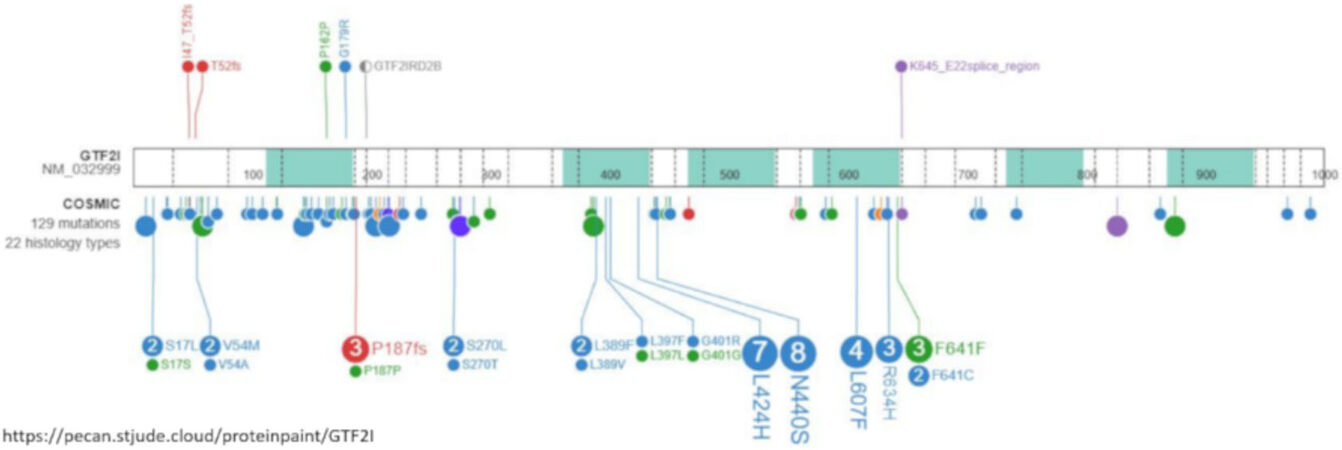
Figure 2. Lollipop plot from St. Judes PECAN database demonstrating somatic point mutations across the GTF2I gene. GTF2I gene (transcript RM_032999) is shown with overlayed data from the COSMIC database. Each individual dot corresponds to a documented mutation. The majority of the mutations are of missense type (blue dots). GTF2I p.L424H is one of the most commonly occurring mutations. Source of data and image: https://pecan.stjude.cloud/ and https://cancer.sanger.ac.uk/cosmic[77-79].
Molecular classification of thymoma
In the past few years, two significant molecular studies have been published that describe potential molecular classification systems for thymomas. The first study by Lee et al.[31] stratified thymomas into four separate molecularly defined groups: (1) the GTF2I group (enriched for GTF2I mutations); (2) the T-cell signaling group characterized by increased expression of genes related to T-cell signaling; (3) the chromosomal stability group characterized by low levels of chromosomal abnormalities, and (4) the chromosomal instability group characterized by increased genetic complexity[31]. Interestingly, these molecular groups roughly correlated with traditional histologic classification and staging parameters: type A and AB thymomas tended to fall into the GTF2I group, type B1 and B2 thymomas tended to fall into the T-cell signaling group, and type B3 thymomas tended to fall into the chromosomal instability group.
A second large, integrated molecular analysis performed by Radovich et al.[37] led to a four-tiered molecular classification system in which thymomas were assigned to one of three molecular subtypes, and thymic carcinoma was assigned to the fourth group. The molecular thymoma subtypes include an A-like cluster and AB-like cluster that share many similarities including frequent GTF2I mutations and overexpression of the C19 miRNA cluster; these two groups are distinguished by differences in regulation of p53, MYB, MYC, and FOXM1. The third molecular thymoma subtype is the B-like cluster characterized by the absence of both GTF2I mutations and overexpression of the C19 miRNA cluster. The B-like subtype is also more closely associated with myasthenia gravis. This study also demonstrated that thymomas generally have low mutational burdens. We recently tested a small cohort of atypical type B3 thymomas using a 50 gene next-generation sequencing panel and similarly found a low number of non-synonymous mutations indicating that even some cases of the more aggressive subtype of thymomas harbor a low number of somatic mutations, although these tumors would benefit from testing with more extensive panels to more clearly delineate this [Figure 3]. A comparison of the aforementioned proposed molecular classification systems is presented in Table 1. More recent studies have identified additional gene expression profiles within different thymoma subsets including increased expression of SOX9[41]. SOX9 expression was positively correlated with multiple other genes including POU2F3 and expression of TRPM5 and KIT[41]. These studies reveal that thymomas show heterogenous molecular alterations that can be corralled into relatively discrete groups. A morpho-molecular classification of thymomas will likely replace the traditional histomorphologic classification systems sometime in the future. This welcomed modification will simplify and improve the classification and prognostication of these lesions. However, at present, no recurrent clinically actionable mutations have been identified, and molecularly guided treatment decisions remain elusive.
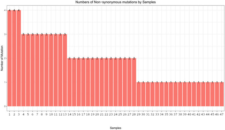
Figure 3. Numbers of non-synonymous mutations in atypical (type B3) thymomas. A bar plot of next-generation sequencing data is shown from 47 atypical thymomas (unpublished data) using the AmpliSeq Cancer Hotspot Panel v2 (Illumina, product number 20019161) using paired-end 2 x 300 read format with an average 500X depth per sample. The bar plot demonstrates a low number of non-synonymous mutations, with the majority of samples showing only two or fewer mutations.
Comparison of proposed molecular classification systems for thymic epithelial neoplasms
| Group | Lee et al. (2017)[31] | Radovich et al. (2018)[34] |
| 1 | GTF2I group | A-like cluster |
| Features: Encompasses type A and AB thymoma, this group is enriched for GTF2I mutations | Features: Corresponds to type A thymoma. GTF2I mutations and C19MC overexpression. Increase in p53 and XBP1-2 expression, decrease in MYC, MYB and FOXM1 expression | |
| 2 | T-cell signaling group | AB-like cluster |
| Features: Encompasses type B1 and B2 thymoma. Characterized by increased expression of genes related T-cell signaling including CD8A, CD8B, CD3D, CTLA4 and others | Features: Corresponds to type AB thymoma, shares some overlap with the A-like group. GTF2I mutations and C19MC overexpression. Increase in MYB, EZF1, and FOXM1 expression, decrease in p53 and Tap73a expression | |
| 3 | Chromosomal stability group | B-like cluster |
| Features: Correlates to type B2 thymomas. This group harbors relatively simple karyotypes with low levels of chromosomal abnormalities | Corresponds to B-type thymomas. Increase in MYC and MYB expression, decrease in p53, XBP1-2, and PPARA_RXRA_coactivator | |
| 4 | Chromosomal instability group | C-like cluster |
| Features: Contains tumors from the B3 group (atypical thymomas), as well as some B2 type thymomas and thymic carcinoma. Characterized by more complex karyotypes with increased chromosomal abnormalities | Features: Corresponds to thymic carcinomas (increased molecular complexity). Chromosome 16q losses, increased MYC and MYB expression, decreased p53, XBP1-2 and PPARA_RXRA_Coactivator expression |
Molecular landscape of thymic carcinoma
Thymic carcinomas represent extremely rare cancers comprising approximately 15%-20% of all TETs. These are separated from their more indolent counterparts based on the presence of aggressive histological features including overt nuclear atypia, necrosis, infiltrative architecture, and loss of the normal organotypical features of the thymus found in thymomas[1,3]. Thymic carcinomas pose a challenge for diagnosis due to a harrowing degree of heterogeneity in their morphology; at least 12 different subtypes of thymic carcinoma have been described to date [Table 2]. Due to their incredibly rare nature, these tumors are difficult to study, and management of these patients may prove difficult in the absence of established molecular targets or consensus treatment guidelines. Limited data exist on the molecular underpinnings of the majority of these tumor types, with only few cases studied to date. In general, thymic carcinomas tend to show increased tumor mutational burdens, multiple copy number alterations, and varied, non-recurrent, somatic mutations[37]. The most prevalent of these tumors is squamous cell carcinoma and variants thereof.
Current WHO classification of thymic carcinoma and associated molecular alterations
| Tumor category | Molecular alterations |
| Squamous cell carcinoma | GTF2I mutations (< 10%) KRAS, NRAS, TP53 mutations Complex chromosomal abnormalities with numerous losses (including homozygous CDKN2A deletions) and gains. Loss of 6q25.2-q25.3 region is seen similar to thymomas Mutations in epigenetic regulatory genes |
| Basaloid carcinoma | Minimal or no data available |
| Lymphoepithelial carcinoma | Various somatic mutations of unknown significance identified EBV-infection seen in up to 50% |
| NUT carcinoma | NUTM1 gene rearrangements; most commonly with BRD3 and BRD4, but many other partner genes reported |
| Clear cell carcinoma | EWSR1-ATF1 rearrangement |
| Mucoepidermoid carcinoma | CRTC1-MAML2 rearrangement |
| Thymic carcinoma with adenoid cystic-like changes | Single case reported with gain of chromosome 8[80] |
| Enteric-type adenocarcinoma | KRAS, TP53, TGFB2 and other somatic point mutations reported |
| Adenocarcinoma, NOS | Minimal or no data available |
| Adenosquamous carcinoma | Minimal or no data available |
| Sarcomatoid carcinoma | Complex chromosomal abnormalities reported |
| Undifferentiated carcinoma | No recurrent or specific abnormalities |
Conventional squamous cell carcinoma has been the most amply studied tumor by molecular techniques. Studies have identified numerous whole and partial arm chromosomal level copy number variations including gains of 1q, 4, 5, 7, 8, 9q, 12, 15, 17q, 18 and 20 and losses of 3p, 6, 6p25, 6q25.2-q25.3, 9p, 13q, 14, 16q, and 17p[21,22,24,27,32,33]. Cases with complex karyotypes have been described[42]. Mutations and methylation of various epigenetic regulatory genes have also been documented including BAP1, ASXL1, SETD2, SMARCA4, TET2, DNMT3A, HOX9, GHSR, and SALL3 have been described[43,44]. Copy number gains of BCL2 and loss of CDKN2A are common[24]. Various somatic point mutations have been documented including RAS family genes, TP53, and only rarely KIT mutations (an unusual finding given that thymic carcinomas commonly overexpress CKIT by immunohistochemistry)[45-50]. GTF2I mutations have only been rarely described in thymic squamous cell carcinomas[37].
Poorly differentiated non-keratinizing thymic squamous cell carcinoma (also referred to as lymphoepithelioma-like carcinoma or lymphoepithelial carcinoma) is a poorly differentiated malignant neoplasm of the thymus characterized by aggressive behavior with poor clinical outcomes. These tumors are associated with Epstein-Barr virus infection in approximately 50% of cases, although tumors in adults tend to be negative for Epstein-Barr Virus[51-53]. The largest molecular study to date identified at least one somatic variant of unknown significance in 16/18 cases[53]. No clinically actionable or targetable mutations were identified; however, the sequencing was performed with a smaller panel of 50 genes. These tumors were also identified to have high levels of PD-L1 expression in a significant number of cases suggesting they may be amenable to immunotherapy regimens[54].
Molecular studies regarding many other types of thymic carcinomas are extremely limited. Many subtypes such as basaloid squamous cell carcinoma, adenosquamous carcinoma, gland forming adenocarcinomas of the thymus, and sarcomatoid carcinomas have either only scattered reports or no testing reported[55-59]. Interestingly, though, rare cases of primary thymic salivary gland tumors appear to show similar molecular alterations to their salivary gland counterparts including EWSR1-ATF1 fusion in clear cell hyalinizing carcinoma and CRTC1-MAML2 fusion in mucoepidermoid carcinoma[60-62]. Finally, although rare, thoracic NUT carcinoma has been relatively well molecularly characterized and is known to harbor a characteristic BRD4-NUTM1 fusion gene in approximately 75% of cases[63-67]. Alternative fusion partners include BRD3 (15% of cases), as well as other less common fusion partners such as NSD3, ZNF532, and ZNF592 (<5% of cases)[66-70].
CONCLUSION
It has only been in the past few decades that the molecular landscape of thymic epithelial neoplasms has begun to be defined. These tumors tend to show a heterogenous complement of molecular alterations, although multi-omic analyses have shown that they can be stratified into different categories based on some recurrent molecular alterations. Of interest are the losses documented at 6q25.2-q25.3 that are found both in thymic carcinoma as well as all subtypes of thymoma, suggesting that these tumors may, in some cases, evolve from pre-existing lower grade lesions; this is corroborated by the fact that thymic carcinomas may rarely be seen arising from thymomas of various subtypes, particularly in combination with atypical (WHO type B3) thymomas[71]. Despite this, many thymic carcinomas arise in the absence of a pre-existing thymoma, and the presence of significant differences in the documented underlying molecular alterations between the two indicates that thymic epithelial cells may arrive at malignancy through more than one molecular pathway. Clinical management and staging of thymomas can be difficult and should be approached in a multidisciplinary setting[72]. The discovery of recurrent alterations including point mutations, fusions, and characteristic micro RNA expressions indicate that these tumors may at some point be amenable to targeted therapies as new treatments are developed. A recent clinical trial (REMORA trial) demonstrated some efficacy with Lenvatinib in patients with advanced thymic carcinoma[73]. Some cases of thymic epithelial neoplasms have also shown alterations that modify cellular signaling pathways such as RAS and mTOR signaling and appear to be amenable to treatments directed at these pathways[74]. Recent studies using immunotherapy have shown conflicting results in aggressive thymic tumors, and additional studies are likely required to identify best use scenarios[75,76]. For now, molecular alterations such as GTF2I mutations or MAML2, EWSR1, and NUT rearrangements, are most valuable in a diagnostic capacity, although rare mutations, such as KIT mutations, may be targeted with specific therapies. Future studies will seek to continue delineating the molecular characteristics of these tumors with the goal of eventually arriving at more precise morpho-molecular classifications that can be used to better guide prognosis, treatment and management of patients with TETs. In current clinical practice, patients with these tumors may benefit from advanced sequencing studies, particularly patients with thymic carcinomas, as the findings can inform the diagnosis and rarely help guide treatment if a clinically actionable mutation is detected.
DECLARATIONS
Authors’ contributionsMade substantial contributions to conception and design of the study and performed data analysis and interpretation: Suster DI, Basu MK, Mackinnon AC
Availability of data and materialsData source: Data included in this manuscript is available upon reasonable request of the corresponding author. Somatic point mutations across the GTF2I gene were obtained from St. Jude Cloud[77-79] (https://www.stjude.cloud).
Financial support and sponsorshipNone.
Conflicts of InterestAll authors declared that there are no conflicts of interest.
Ethical approval and consent to participateNot applicable.
Consent for publicationNot applicable.
Copyright© The Author(s) 2022.
REFERENCES
1. . WHO. Classification of tumors editorial board. Thoracic tumours, 5th ed.; vol. 5. Lyon: IARC Press; 2021.
3. Suster D, Mino-Kenudson M, Suster S. Diagnostic pathology: thoracic, 5th ed.; Philadelphia, PA: Eslevier; 2022.
4. Detterbeck FC, Stratton K, Giroux D, et al. The IASLC/ITMIG thymic epithelial tumors staging project: proposal for an evidence-based stage classification system for the forthcoming (8th) edition of the TNM classification of malignant tumors. J Thorac Oncol 2014;9:S65-72.
5. Filosso PL, Ruffini E, Lausi PO, et al. Historical perspectives: the evolution of the thymic epithelial tumors staging system. Lung Cancer 2014;83:126-32.
6. Weissferdt A, Kalhor N, Bishop JA, et al. Thymoma: a clinicopathological correlation of 1470 cases. Hum Pathol 2018;73:7-15.
7. Roden AC, Yi ES, Jenkins SM, et al. Reproducibility of 3 histologic classifications and 3 staging systems for thymic epithelial neoplasms and its effect on prognosis. Am J Surg Pathol 2015;39:427-41.
8. Moran CA, Walsh G, Suster S, Kaiser L. Thymomas II: a clinicopathologic correlation of 250 cases with a proposed staging system with emphasis on pathologic assessment. Am J Clin Pathol 2012;137:451-61.
9. Suster S, Moran CA. Problem areas and inconsistencies in the WHO classification of thymoma. Semin Diagn Pathol 2005;22:188-97.
10. Detterbeck FC, Marom EM. Thymus. In: Amin MB, Greene FL, Edge SB, et al., editors. AJCC Cancer Staging Manual. Chicago, IL: Springer; 2017. p. 423-29.
11. Koga K, Matsuno Y, Noguchi M, et al. A review of 79 thymomas: modification of staging system and reappraisal of conventional division into invasive and non-invasive thymoma. Pathol Int 1994;44:359-67.
12. Masaoka A, Monden Y, Nakahara K, Tanioka T. Follow-up study of thymomas with special reference to their clinical stages. Cancer 1981;48:2485-92.
13. Verghese ET, den Bakker MA, Campbell A, et al. Interobserver variation in the classification of thymic tumours--a multicentre study using the WHO classification system. Histopathology 2008;53:218-23.
14. Hsu CH, Chan JK, Yin CH, Lee CC, Chern CU, Liao CI. Trends in the incidence of thymoma, thymic carcinoma, and thymic neuroendocrine tumor in the United States. PLoS ONE 2019;14:e0227197.
15. Jong WK, Blaauwgeers JL, Schaapveld M, Timens W, Klinkenberg TJ, Groen HJ. Thymic epithelial tumours: a population-based study of the incidence, diagnostic procedures and therapy. Eur J Cancer 2008;44:123-30.
16. Weis CA, Yao X, Deng Y, et al. Contributors to the ITMIG Retrospective Database. The impact of thymoma histotype on prognosis in a worldwide database. J Thorac Oncol 2015;10:367-72.
17. Marx A, Willcox N, Leite MI, et al. Thymoma and paraneoplastic myasthenia gravis. Autoimmunity 2010;43:413-27.
18. Zaman M, Huissoon A, Buckland M, et al. Clinical and laboratory features of seventy-eight UK patients with Good's syndrome (thymoma and hypogammaglobulinaemia). Clin Exp Immunol 2019;195:132-8.
19. Kisand K, Bøe Wolff AS, Podkrajsek KT, et al. Chronic mucocutaneous candidiasis in APECED or thymoma patients correlates with autoimmunity to Th17-associated cytokines. J Exp Med 2010;207:299-308.
20. Christopoulos P, Dopfer EP, Malkovsky M, et al. A novel thymoma-associated immunodeficiency with increased naive T cells and reduced CD247 expression. J Immunol 2015;194:3045-53.
21. Inoue M, Starostik P, Zettl A, et al. Correlating genetic aberrations with World Health Organization-defined histology and stage across the spectrum of thymomas. Cancer Res 2003;63:3708-15.
22. Penzel R, Hoegel J, Schmitz W, et al. Clusters of chromosomal imbalances in thymic epithelial tumours are associated with the WHO classification and the staging system according to Masaoka. Int J Cancer 2003;105:494-8.
23. Petrini I, Rajan A, Pham T, et al. Whole genome and transcriptome sequencing of a B3 thymoma. PLoS ONE 2013;8:e60572.
24. Petrini I, Meltzer PS, Zucali PA, et al. Copy number aberrations of BCL2 and CDKN2A/B identified by array-CGH in thymic epithelial tumors. Cell Death Dis 2012;3:e351.
25. Petrini I, Wang Y, Zucali PA, et al. Copy number aberrations of genes regulating normal thymus development in thymic epithelial tumors. Clin Cancer Res 2013;19:1960-71.
26. Cin P, De Wolf-peeters C, Deneffe G, Fryns J, Van den Berghe H. Thymoma with a t(15;22)(p11;q11). Cancer Genet Cytogenet 1996;89:181-3.
27. Zettl A, Ströbel P, Wagner K, et al. Recurrent genetic aberrations in thymoma and thymic carcinoma. Am J Pathol 2000;157:257-66.
28. Kristoffersson U, Heim S, Mandahl N, Åkerman M, Mitelman F. Multiple clonal chromosome aberrations in two thymomas. Cancer Genet Cytogenet 1989;41:93-8.
29. Mirza I, Kazimi SN, Ligi R, Burns J, Braza F. Cytogenetic profile of a thymoma. A case report and review of the literature. Arch Pathol Lab Med 2000;124:1714-6.
30. den Berghe I, Debiec-rychter M, Proot L, Hagemeijer A, Michielssen P. Ring chromosome 6 may represent a cytogenetic subgroup in benign thymoma. Cancer Genet Cytogenet 2002;137:75-7.
31. Lee GY, Yang WI, Jeung HC, et al. Genome-wide genetic aberrations of thymoma using cDNA microarray based comparative genomic hybridization. BMC Genomics 2007;8:305.
32. Girard N, Shen R, Guo T, et al. Comprehensive genomic analysis reveals clinically relevant molecular distinctions between thymic carcinomas and thymomas. Clin Cancer Res 2009;15:6790-9.
33. Enkner F, Pichlhöfer B, Zaharie AT, et al. Molecular profiling of thymoma and thymic carcinoma: genetic differences and potential novel therapeutic targets. Pathol Oncol Res 2017;23:551-64.
34. Radovich M, Solzak JP, Hancock BA, et al. A large microRNA cluster on chromosome 19 is a transcriptional hallmark of WHO type A and AB thymomas. Br J Cancer 2016;114:477-84.
35. Petrini I, Meltzer PS, Kim IK, et al. A specific missense mutation in GTF2I occurs at high frequency in thymic epithelial tumors. Nat Genet 2014;46:844-9.
36. Feng Y, Lei Y, Wu X, et al. GTF2I mutation frequently occurs in more indolent thymic epithelial tumors and predicts better prognosis. Lung Cancer 2017;110:48-52.
37. Radovich M, Pickering CR, Felau I, et al. Cancer genome atlas network. the integrated genomic landscape of thymic epithelial tumors. Cancer Cell 2018;33:244-258.e10.
38. Vivero M, Davineni P, Nardi V, Chan JKC, Sholl LM. Metaplastic thymoma: a distinctive thymic neoplasm characterized by YAP1-MAML2 gene fusions. Mod Pathol 2020;33:560-5.
40. Massoth LR, Hung YP, Dias-Santagata D, et al. Pan-Cancer landscape analysis reveals recurrent. KMT2A ;4:PO.19.00288.
41. Sonobe H, Ohtsuki Y, Nakayama H, Asaba K, Nishiya K, Shimizu K. A thymic squamous cell carcinoma with complex chromosome abnormalities. Cancer Genet Cytogenet 1998;103:83-5.
42. Yuan X, Huang L, Luo W, et al. Diagnostic and prognostic significances of SOX9 in thymic epithelial tumor. Front Oncol 2021;11:708735.
43. Wang Y, Thomas A, Lau C, et al. Mutations of epigenetic regulatory genes are common in thymic carcinomas. Sci Rep 2014;4:7336.
44. Kishibuchi R, Kondo K, Soejima S, et al. DNA methylation of GHSR, GNG4, HOXD9 and SALL3 is a common epigenetic alteration in thymic carcinoma. Int J Oncol 2020;56:315-26.
45. Yoh K, Nishiwaki Y, Ishii G, et al. Mutational status of EGFR and KIT in thymoma and thymic carcinoma. Lung Cancer 2008;62:316-20.
46. Tsuchida M, Umezu H, Hashimoto T, et al. Absence of gene mutations in KIT-positive thymic epithelial tumors. Lung Cancer 2008;62:321-5.
47. Petrini I, Zucali PA, Lee HS, et al. Expression and mutational status of c-kit in thymic epithelial tumors. J Thorac Oncol 2010;5:1447-53.
48. Hirabayashi H, Fujii Y, Sakaguchi M, et al. p16INK4, pRB, p53 and cyclin D1 expression and hypermethylation ofCDKN2 gene in thymoma and thymic carcinoma. Int J Cancer 1997;73:639-44.
49. Tateyama H, Eimoto T, Tada T, et al. p53 protein expression and p53 gene mutation in thymic epithelial tumors. An immunohistochemical and DNA sequencing study. Am J Clin Pathol 1995;104:375-81.
50. Sakane T, Murase T, Okuda K, et al. A mutation analysis of the EGFR pathway genes, RAS, EGFR, PIK3CA, AKT1 and BRAF, and TP53 gene in thymic carcinoma and thymoma type A/B3. Histopathology 2019;75:755-66.
51. Hsueh C, Kuo TT, Tsang NM, Wu YC, Yang CP, Hung IJ. Thymic lymphoepitheliomalike carcinoma in children: clinicopathologic features and molecular analysis. J Pediatr Hematol Oncol 2006;28:785-90.
52. Chen PC, Pan CC, Yang AH, Wang LS, Chiang H. Detection of Epstein-Barr virus genome within thymic epithelial tumours in Taiwanese patients by nested PCR, PCR in situ hybridization, and RNA in situ hybridization. J Pathol 2002;197:684-8.
53. Suster D, Pihan G, Mackinnon AC, Suster S. Poorly Differentiated nonkeratinizing squamous cell carcinoma of the thymus: clinicopathologic and molecular genetic study of 25 cases. Am J Surg Pathol 2018;42:1224-36.
54. Suster D, Pihan G, Mackinnon AC, Suster S. Expression of PD-L1/PD-1 in lymphoepithelioma-like carcinoma of the thymus. Mod Pathol 2018;31:1801-6.
56. Lee Y, Park S, Lee SH, Lee H. Characterization of genetic aberrations in a single case of metastatic thymic adenocarcinoma. BMC Cancer 2017;17:330.
57. Maghbool M, Ramzi M, Nagel I, et al. Primary adenocarcinoma of the thymus: an immunohistochemical and molecular study with review of the literature. BMC Clin Pathol 2013;13:17.
58. Eimoto T, Kitaoka M, Ogawa H, et al. Thymic sarcomatoid carcinoma with skeletal muscle differentiation: report of two cases, one with cytogenetic analysis. Histopathology 2002;40:46-57.
59. Porubsky S, Jessup P, Kee D, et al. Potentially actionable FGFR2 high-level amplification in thymic sebaceous carcinoma. Virchows Arch 2020;476:323-7.
60. Porubsky S, Rudolph B, Rückert JC, et al. International Thymic Malignancy Interest Group (ITMIG). EWSR1 translocation in primary hyalinising clear cell carcinoma of the thymus. Histopathology 2019;75:431-6.
61. Roden AC, Erickson-Johnson MR, Yi ES, García JJ. Analysis of MAML2 rearrangement in mucoepidermoid carcinoma of the thymus. Hum Pathol 2013;44:2799-805.
62. Prieto-Granada CN, Inagaki H, Mueller J. Thymic mucoepidermoid carcinoma: report of a case with CTRC1/3-MALM2 molecular studies. Int J Surg Pathol 2015;23:277-83.
63. Evans AG, French CA, Cameron MJ, et al. Pathologic characteristics of NUT midline carcinoma arising in the mediastinum. Am J Surg Pathol 2012;36:1222-7.
64. Gökmen-Polar Y, Kesler K, Loehrer PJ Sr, Badve S. NUT midline carcinoma masquerading as a thymic carcinoma. J Clin Oncol 2016;34:e126-9.
65. Gökmen-Polar Y, Cano OD, Kesler KA, Loehrer PJ, Badve S. NUT midline carcinomas in the thymic region. Mod Pathol 2014;27:1649-56.
66. French CA, Miyoshi I, Kubonishi I, et al. BRD4-NUT fusion oncogene: a novel mechanism in aggressive carcinoma. Cancer Res 2003;63:304-7.
67. French CA, Ramirez CL, Kolmakova J, et al. BRD-NUT oncoproteins: a family of closely related nuclear proteins that block epithelial differentiation and maintain the growth of carcinoma cells. Oncogene 2008;27:2237-42.
68. Shiota H, Elya JE, Alekseyenko AA, et al. “Z4” Complex member fusions in NUT carcinoma: implications for a novel oncogenic mechanism. Mol Cancer Res 2018;16:1826-33.
69. Alekseyenko AA, Walsh EM, Zee BM, et al. Ectopic protein interactions within BRD4-chromatin complexes drive oncogenic megadomain formation in NUT midline carcinoma. Proc Natl Acad Sci USA 2017;114:E4184-92.
70. French CA, Rahman S, Walsh EM, et al. NSD3-NUT fusion oncoprotein in NUT midline carcinoma: implications for a novel oncogenic mechanism. Cancer Discov 2014;4:928-41.
71. Suster DI, Craig Mackinnon A, DiStasio M, Basu MK, Pihan G, Suster S. Atypical thymomas with squamoid and spindle cell features: clinicopathologic, immunohistochemical and molecular genetic study of 120 cases with long-term follow-up. Mod Pathol 2022; doi: 10.1038/s41379-022-01013-x.
72. Hamaji M, Ali SO, Burt BM. A meta-analysis of induction therapy for advanced thymic epithelial tumors. Ann Thorac Surg 2015;99:1848-56.
73. Sato J, Satouchi M, Itoh S, et al. Lenvatinib in patients with advanced or metastatic thymic carcinoma(REMORA): a multicentre, phase 2 trial. Lancet Oncol 2020;21:843-50.
74. Wheler J, Hong D, Swisher SG, et al. Thymoma patients treated in a phase I clinic at MD Anderson Cancer Center: responses to mTOR inhibitors and molecular analyses. Oncotarget 2013;4:890-8.
75. Giaccone G, Kim C, Thompson J, et al. Pembrolizumab in patients with thymic carcinoma: a single-arm, single-centre, phase 2 study. Lancet Oncol 2018;19:347-55.
76. Katsuya Y, Horinouchi H, Seto T, et al. Single-arm, multicentre, phase II trial of nivolumab for unresectable or recurrent thymic carcinoma: PRIMER study. Eur J Cancer 2019;113:78-86.
77. McLeod C, Gout AM, Zhou X, et al. St. Jude cloud: a pediatric cancer genomic data-sharing ecosystem. Cancer Discov 2021;11:1082-99.
78. Edmonson MN, Patel AN, Hedges DJ, et al. Pediatric Cancer Variant Pathogenicity Information Exchange (PeCanPIE): a cloud-based platform for curating and classifying germline variants. Genome Res 2019;29:1555-65.
79. Zhou X, Wang J, Patel J, et al. Exploration of coding and non-coding variants in cancer using genomepaint. Cancer Cell 2021;39:83-95.e4.
Cite This Article
Export citation file: BibTeX | RIS
OAE Style
Suster DI, Basu MK, Mackinnon AC. Molecular pathology of thymoma and thymic carcinoma. J Cancer Metastasis Treat 2022;8:19. http://dx.doi.org/10.20517/2394-4722.2021.210
AMA Style
Suster DI, Basu MK, Mackinnon AC. Molecular pathology of thymoma and thymic carcinoma. Journal of Cancer Metastasis and Treatment. 2022; 8(5): 19. http://dx.doi.org/10.20517/2394-4722.2021.210
Chicago/Turabian Style
Suster, David I., Malay Kumar Basu, A. Craig Mackinnon. 2022. "Molecular pathology of thymoma and thymic carcinoma" Journal of Cancer Metastasis and Treatment. 8, no.5: 19. http://dx.doi.org/10.20517/2394-4722.2021.210
ACS Style
Suster, DI.; Basu MK.; Mackinnon AC. Molecular pathology of thymoma and thymic carcinoma. J. Cancer. Metastasis. Treat. 2022, 8, 19. http://dx.doi.org/10.20517/2394-4722.2021.210
About This Article
Special Issue
Copyright

Data & Comments
Data


 Cite This Article 20 clicks

 Like This Article 32
likes
Like This Article 32
likes


Comments
Comments must be written in English. Spam, offensive content, impersonation, and private information will not be permitted. If any comment is reported and identified as inappropriate content by OAE staff, the comment will be removed without notice. If you have any queries or need any help, please contact us at support@oaepublish.com.